Endodontics - what it is, the 5 signs that mean you need it, and what treatment actually involves
Most patients have never heard the word endodontics until a dentist uses it in an appointment. At that point, they know only two things: it is something clinical, and it sounds serious.
Endodontics is the branch of dentistry concerned with the dental pulp and the periapical tissues: the structures inside the tooth and at the tip of the root. It covers the diagnosis, treatment and prevention of diseases affecting these structures. In practical terms, this primarily means root canal treatment, and it also includes retreatment of failed root canals and surgical procedures at the root tip.
It is not an especially dramatic branch of dentistry. What it does is save teeth. The alternative to successful endodontic treatment is extraction, followed by the considerably more complex, time-consuming and costly process of replacing what has been lost. Understanding what endodontics involves, what the signs of needing it are, and what treatment actually looks like removes a lot of the anxiety that tends to accompany the recommendation.
At Smile Perfections in Oadby, Leicester, led by Dr Juttes Pallipatt GDC No. 104499 and Dr Pratima Pallipatt GDC No. 101258, root canal treatment is carried out with current techniques and materials, and we take the time to ensure every patient understands what treatment involves before any procedure begins.
The anatomy endodontics works with
Before explaining what endodontics treats, it is worth understanding the structures it deals with, because the anatomy explains everything about why these tissues become diseased and how treatment works.
The dental pulp
Every tooth contains a central chamber of soft tissue called the dental pulp. This tissue consists of blood vessels, lymphatic vessels, nerve fibres and connective tissue. It is surrounded on all sides by dentine, the hard mineralised tissue that makes up the bulk of the tooth.
The pulp occupies the pulp chamber in the crown of the tooth and extends down into one or more root canals that run through the length of each root. The canal tapers toward the root apex (tip) and exits at the apical foramen, a small opening through which the blood vessels and nerves enter and exit the tooth.
The dental pulp plays an important role during tooth development: it produces the dentine that forms the tooth and provides the sensation that allows the tooth to respond to stimuli. Once the tooth is fully developed, the pulp’s role is less essential, which is why a tooth that has had its pulp removed can still function for many years.
The periapical tissues
Around the apex of each root lies the periapical area: the bone, the periodontal ligament fibers and the connective tissue at the root tip. When the pulp becomes infected, the infection spreads through the root canal system and exits at the apex, causing inflammation and infection of this periapical tissue. This is periapical periodontitis, and it is what produces the abscess and bone destruction associated with untreated pulp infection.
Why endodontic treatment becomes necessary
The pulp can become diseased through two main pathways: progressive decay reaching the pulp, and trauma to the tooth.
Decay reaching the pulp
The enamel is the hardest substance in the body, but it is not immune to the acid produced by plaque bacteria. Once decay penetrates the enamel and enters the dentine, it progresses faster because dentine is less mineralised and more porous. A cavity that is not treated grows, and a growing cavity approaches the pulp chamber.
When the cavity is still confined to the outer dentine, the pulp responds with reversible inflammation: a temporary heightened sensitivity that resolves once the decay is removed and the tooth is restored. When the decay reaches within approximately 1mm of the pulp, or breaches it, the response becomes irreversible. The pulp cannot recover from this degree of insult. The inflammatory process progresses to pulp necrosis (death of the pulp tissue) and, without treatment, to infection of the periapical tissues.
Trauma and cracking
Physical trauma to a tooth, whether from a fall, a sports injury or a bite on something unexpectedly hard, can damage the pulp without necessarily causing visible crown damage. A tooth that receives a significant blow may have its blood supply disrupted. The pulp, deprived of blood supply, undergoes necrosis over weeks to months. This is why a tooth involved in trauma is monitored over time even if it looks intact immediately after the incident.
Cracks in the tooth allow bacteria to penetrate to the pulp along the crack line, causing infection. Deep cracks that reach the pulp invariably require endodontic treatment, and if they extend below the gum line into the root, the tooth may not be salvageable.
The 5 signs that endodontic treatment might be needed
This is where clinical signs and patient experience meet. These are the presentations that most commonly lead to an endodontic assessment at a dental check-up.
Sign 1: Spontaneous pain that is not triggered by anything
Pain that starts without any stimulus, arrives unexpectedly, and persists after the trigger (if any) is removed is a cardinal symptom of irreversible pulpitis. Unlike the sensitivity of dentine hypersensitivity, which is brief and stimulus-dependent, the pain of irreversible pulpitis is often prolonged, may be throbbing, and can wake patients from sleep.
Sign 2: Severe sensitivity to heat that lingers
Cold sensitivity that lingers for more than 30 seconds after the cold stimulus is removed is a consistent clinical indicator of irreversible pulpitis. Even more specific: heat sensitivity that gets worse rather than better once the heat is removed suggests a pulp that has reached advanced inflammatory change. Some patients with irreversible pulpitis specifically seek out cold drinks because they provide temporary relief from spontaneous heat pain.
Sign 3: A tooth that feels raised or tender to bite on
When the periapical tissues become inflamed, the slight movement that every tooth has within its socket becomes painful. The tooth feels as though it is raised higher than the surrounding teeth. The pressure of biting on that tooth is uncomfortable to the point of being difficult to tolerate. This periapical tenderness is a clinical sign that the infection has extended beyond the root apex.
Sign 4: A visible swelling or a bad taste
A gum boil (parulis) on the gum alongside a tooth, or a persistent bad taste from one area of the mouth, often indicates a sinus tract: a channel through which pus from a periapical abscess is draining. The swelling may look like a small pimple on the gum and may periodically discharge, temporarily reducing the pain before building again. This is not a sign the problem is resolving; it is a sign the infection is chronic and established.
Sign 5: A tooth that has changed colour
A tooth that has gradually become darker than its neighbours, typically taking on a grey or brownish hue, has usually had a pulp that has died and begun to degrade internally. Blood products from the dying pulp tissue seep into the dentinal tubules and produce the characteristic discolouration. This can happen with no pain at all if the nerve has died gradually and without acute infection.
If you have noticed any of these signs, the appropriate first step is a dental check-up with appropriate diagnostic testing to establish what is happening inside the tooth.
What endodontic treatment involves: the clinical sequence
Root canal treatment has a reputation that significantly outpaces the reality of modern technique. Understanding the actual procedure removes much of the apprehension.
Diagnosis and planning
Before any treatment begins, the tooth is assessed clinically and radiographically. Thermal tests (using cold) and electric pulp testing establish whether the pulp is vital, partially vital or non-vital. Percussion testing identifies periapical tenderness. A periapical X-ray shows the root anatomy, the number and curvature of the canals, and any periapical pathology. In complex cases, a cone beam CT scan (CBCT) gives a three-dimensional view of the root canal system that a flat X-ray cannot provide.
Access and canal identification
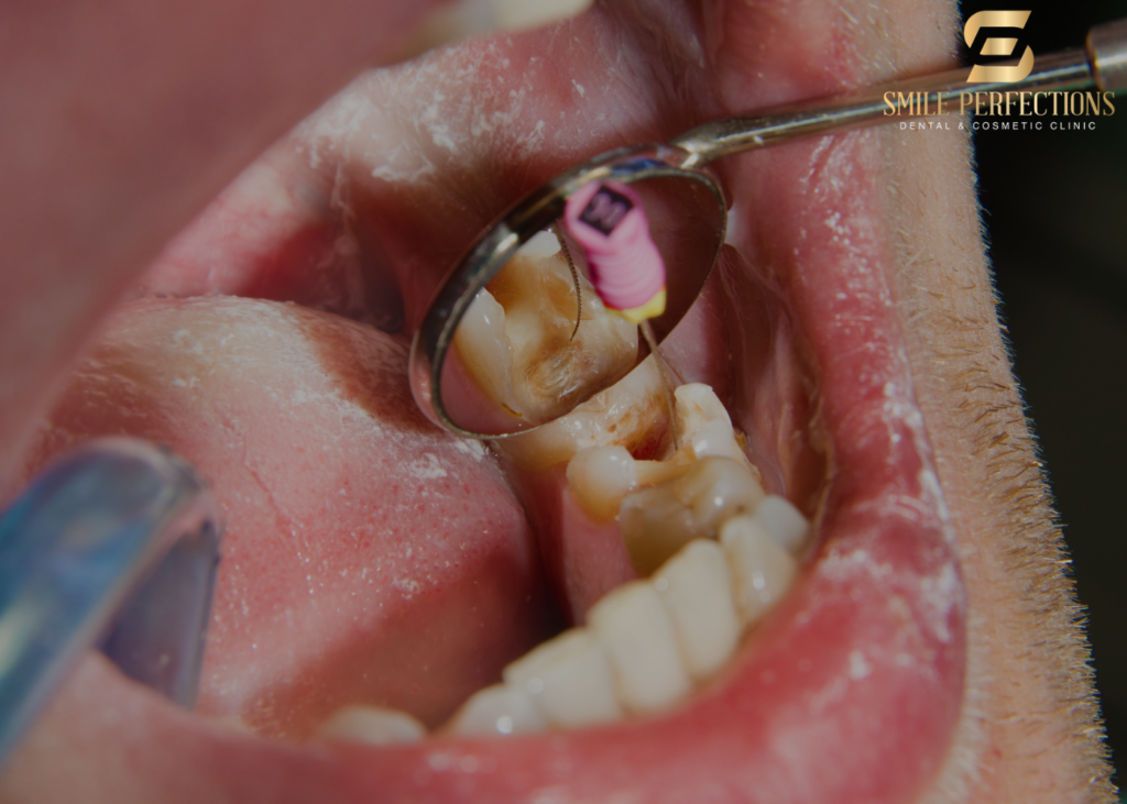
Under local anaesthetic, and typically with rubber dam isolation (a thin sheet of rubber that isolates the tooth from the rest of the mouth, keeping it clean and dry), an access cavity is created through the crown of the tooth. This allows direct access to the pulp chamber. The canal orifices are identified and their number established.
Teeth are more anatomically complex internally than they appear externally. Lower molar teeth have two or three roots, each containing one to three canals. Upper molars typically have three roots with three to four canals. This anatomical variation is why endodontic treatment can vary in complexity considerably between tooth types.
Canal preparation
The root canal system is prepared using a combination of hand files and rotary nickel-titanium instruments. These are used sequentially to shape and clean the canals, enlarging them from the original diameter while preserving the natural curvature of the root. The goal is a tapered preparation that allows thorough irrigation and later filling.
Throughout preparation, copious irrigation with sodium hypochlorite (NaOCl) and EDTA solutions dissolves organic tissue remnants, disrupts the bacterial biofilm within the canal, and removes the smear layer (a debris layer left by instrumentation) to allow the irrigants and final sealer to penetrate the dentinal tubules.
Obturation (filling the canal system)
Once the canals are clean and shaped, they are filled with gutta-percha (a natural thermoplastic material) and a sealer to create a three-dimensional, hermetically sealed fill of the entire canal system, including lateral canals and the apical foramen. The quality of this seal is one of the most significant determinants of long-term success.
Restoration
A root canal treated tooth needs a definitive restoration. Because root canal treatment removes the pulp tissue that previously provided fluid to the dentine, the tooth becomes more brittle over time. For posterior teeth (molars and premolars), a crown is typically the recommended restoration to prevent cuspal fracture, which would otherwise be the most common cause of failure after successful endodontic treatment. This is discussed and planned at the same time as the endodontic treatment.
How modern endodontics has changed the experience
The generation of patients whose memory of dentistry includes the phrase “root canal” as a byword for suffering had their experiences shaped by older technology and older anaesthetic techniques. Modern endodontics is a different proposition.
Nickel-titanium rotary systems have replaced the hand-filing that was both time-consuming and less predictable. Rotary instruments follow the natural curvature of the canal with considerably less risk of straightening or perforating it.
CBCT imaging provides three-dimensional anatomical information that allows complex cases with unusual root anatomy to be planned and executed with a level of precision that was previously impossible.
Modern local anaesthetics, including articaine with its excellent bone penetration, combined with specific techniques such as intraosseous injection for difficult-to-numb lower molars, means that patients with acutely inflamed teeth, which were historically the most challenging to anaesthetise, can now be made reliably comfortable throughout the procedure.
Electronic apex locators determine the precise length of each root canal within a fraction of a millimetre, replacing the more approximate radiographic estimation of canal length.
Magnification and illumination, using surgical loupes or dental operating microscopes, allow the clinician to identify canal orifices, detect cracks and assess the quality of the preparation at a level invisible to the naked eye.
The honest clinical truth: root canal treatment carried out with modern technique, by an experienced clinician, is no more uncomfortable during the procedure than a filling. The discomfort that patients experience is almost universally the pain before treatment, not during it.
Success rates and what determines them
Endodontic treatment has excellent success rates when carried out well and when the tooth is appropriately restored afterwards. The published literature consistently reports success rates above 90% at five-year follow-up for initial root canal treatment in teeth without periapical pathology, and 80 to 85% for teeth with established periapical lesions at the time of treatment.
The main factors that affect success are:
- The quality of the canal preparation and obturation. A poorly shaped canal cannot be cleaned effectively. An inadequate seal allows reinfection. The technical quality of the procedure is the most controllable factor in success.
- The quality of the coronal restoration. Studies consistently show that a well-executed root canal followed by a poor restoration fails more often than a technically average root canal followed by an excellent crown. The restoration seals the access cavity and protects the tooth from reinfection and fracture.
- Pre-treatment periapical pathology. Teeth with established periapical abscesses have a slightly lower success rate than those without, because the bone destruction provides a reservoir for bacteria that the root canal treatment must eliminate. This is one of the reasons treating teeth earlier, before abscess formation, produces better outcomes.
- Patient factors. Diabetes, smoking and compromised immune function are associated with reduced healing capacity and slightly lower success rates.
When initial root canal treatment has failed, endodontic retreatment is the appropriate next step before extraction is considered. Retreatment involves removing the existing filling, re-negotiating the canal system, and repeating the procedure. Where retreatment is not appropriate or has not succeeded, surgical endodontics (apicoectomy) removes the infected root tip and seals the apex from the outside, through a small incision in the gum.
The role of oral hygiene in protecting endodontically treated teeth
A tooth that has had root canal treatment can still develop new decay and gum disease around it. The absence of a nerve does not make the tooth immune to the same processes that affect any other tooth. Gum disease that destroys the bone around a root-treated tooth affects the tooth just as it would any other.
Regular dental hygienist appointments maintain the gum health around treated teeth, remove the bacterial deposits that would otherwise drive gum disease, and allow early identification of any new decay at the crown margin. Consistent professional maintenance extends the functional lifespan of any restoration, including crowns placed following endodontic treatment.
In conclusion
Endodontics is the branch of dentistry that saves teeth from the inside. When the pulp becomes inflamed beyond recovery or infected, root canal treatment removes the source of the problem, cleans the canal system, seals it against reinfection, and allows the tooth to be restored to full function. The alternative is extraction and the more complex and costly process of replacement.
The five signs that endodontic treatment may be needed are: spontaneous pain, heat sensitivity that lingers, pain on biting, a draining abscess, and a tooth that has changed colour. Any of these warrants a dental check-up with appropriate diagnostic testing.
At Smile Perfections in Oadby, Leicester, Dr Juttes Pallipatt GDC No. 104499 and Dr Pratima Pallipatt GDC No. 101258 provide thorough endodontic assessment and treatment with the clinical precision that the procedure demands.
Patients frequently ask
The duration depends on the tooth involved and the complexity of the root canal system. An upper incisor with a single, straight root canal may be completed in one appointment of approximately 60 to 90 minutes. A lower molar with three roots and potentially four canals is a more extended procedure, often requiring two appointments of 90 minutes each. Modern techniques, including rotary instrumentation and electronic apex locators, have significantly reduced treatment times compared to fully manual approaches.
The procedure is carried out under local anaesthetic. With modern anaesthetic agents and technique, including the intraosseous injection where standard infiltration is insufficient, the procedure should involve pressure and movement but not pain. The pain patients associate with root canal treatment is almost always the acute pulpitis or abscess pain before the procedure, not the procedure itself. Post-operative soreness for one to three days after treatment is common and manageable with over-the-counter ibuprofen and paracetamol.
An infected pulp does not recover without clinical treatment. The infection progresses from the pulp through the root canal system into the periapical bone, causing bone destruction and abscess formation. The pain typically worsens. Facial swelling may develop. In rare but documented cases, dental infection can spread into the spaces of the neck and, if untreated, become life-threatening. Extraction ultimately resolves the infection but requires replacing the missing tooth. The sooner endodontic treatment is carried out, the simpler the treatment and the better the prognosis for the tooth.
Yes, in principle. A well-treated tooth with a quality restoration, maintained with good oral hygiene and regular hygienist appointments, can last as long as any natural tooth. The factors most commonly responsible for eventual failure are crown fracture (which is why a crown rather than just a filling is recommended for posterior teeth), new decay at the crown margin, and gum disease around the tooth. All of these are preventable with appropriate maintenance.
An endodontist is a dental specialist who has completed additional postgraduate training specifically in endodontics, typically two to three years beyond general dental qualification. Endodontists manage the most complex cases, including retreatment, surgical endodontics and cases with unusual root anatomy or calcified canals. General dentists with appropriate experience and training carry out root canal treatment routinely and successfully for straightforward to moderately complex cases. At Smile Perfections, the clinical assessment of each case determines whether treatment is appropriate to carry out in practice or whether specialist referral would serve the patient better.
Medical and dental information disclaimer
The information in this article is intended for general educational guidance only and does not constitute personalised dental advice. For concerns about tooth pain, sensitivity or suspected pulp pathology, please book an appointment with a qualified dental professional for a proper clinical assessment.
Smile Perfections is a private dental practice in Oadby, Leicester, led by Dr Juttes Pallipatt GDC No. 104499 and Dr Pratima Pallipatt GDC No. 101258. We offer root canal treatment, dental check-ups, dental hygienist appointments, Invisalign, composite bonding, porcelain veneers, teeth whitening, dental crowns and smile makeovers.