Tooth anatomy explained
Most people live their entire lives without giving much thought to what their teeth are actually made of. They are just there: hard, white, useful for eating, and occasionally sources of pain. But the internal structure of a tooth is considerably more complex than a block of hard material, and understanding that structure explains almost everything about how decay works, why fillings fail, what root canal treatment does, why gum disease leads to tooth loss, and why certain dental treatments produce the results they do.
Tooth anatomy explained properly is a foundation for understanding your own dental health, and for making sense of what a dentist is actually doing when they treat any given condition.
At Smile Perfections in Oadby, Leicester, led by Dr Juttes Pallipatt GDC No. 104499 and Dr Pratima Pallipatt GDC No. 101258, the clinical work carried out on every patient is shaped directly by tooth anatomy and the behaviour of each tissue under different conditions. Here is the complete picture.
The basic divisions of a tooth
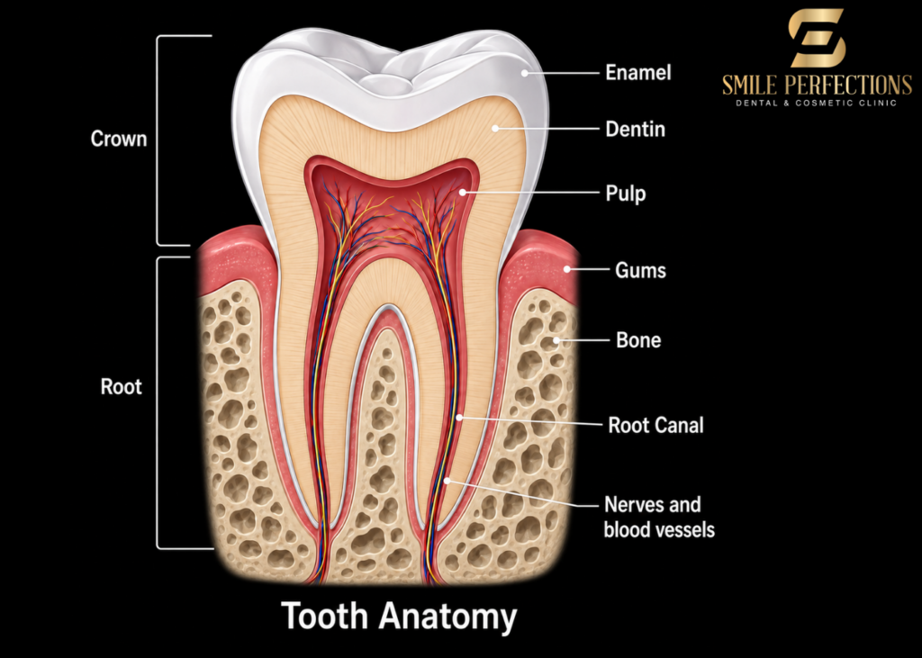
Before getting into the individual tissues, it helps to understand the basic structural divisions that every tooth shares.
The crown: The visible portion of the tooth above the gum line. The anatomical crown is the full extent of the enamel-covered portion; the clinical crown is whatever portion is visible in the mouth at a given time (these differ in gum recession, where more than the anatomical crown may be visible, or in altered passive eruption, where gum tissue covers part of the anatomical crown).
The root: The portion of the tooth embedded in the jawbone. Roots vary considerably in number and shape between tooth types. Single-rooted teeth (incisors, canines, most premolars) have one root. Molars have two (lower) or three (upper) roots. Root length, curvature and anatomy are clinically significant for extractions, root canal treatment and implant planning.
The cervix (neck): The junction between the crown and root, at the level of the gum line. This is called the cemento-enamel junction (CEJ), where the enamel of the crown meets the cementum of the root. The CEJ is an important anatomical landmark in periodontal assessment and in restorative planning.
Enamel: the outer armour of the crown
Enamel is the hardest biological substance in the human body. With a Vickers hardness of approximately 250 to 390 (compared to dentine at around 68), it is significantly harder than bone and considerably harder than any other bodily tissue. This hardness comes from its extraordinary degree of mineralisation.
What enamel is made of: Approximately 96% inorganic mineral by weight, primarily in the form of carbonated hydroxyapatite (Ca₁₀(PO₄)₆(OH)₂), arranged in highly organised crystalline rods called enamel prisms (or enamel rods). Each prism runs from the dentine-enamel junction to the outer enamel surface. The remaining 4% is water and organic material, including enamel proteins from the ameloblasts (the cells that produced the enamel during development).
Critical fact about enamel: Once formed, enamel cannot be regenerated by the body. Ameloblasts are lost when the tooth erupts. The body can remineralise early enamel lesions (adding mineral back into partially demineralised enamel) with fluoride and salivary calcium and phosphate, but cannot produce new enamel to replace what has been lost to decay or acid erosion. This is tooth anatomy explained at its most clinically consequential: every bit of enamel lost is permanently lost.
Why enamel looks the way it does: Enamel is actually translucent rather than opaque white. The white or cream colour of teeth comes primarily from the dentine beneath, which shows through the translucent enamel. Enamel thickness varies across the crown, reaching 2 to 2.5mm at the cusps and incisal edges and becoming very thin toward the cervix. This explains why teeth appear more translucent and slightly grey-blue at the biting edges: the enamel is thicker there, so the dentine colour contributes less.
What this means for dental treatment: Teeth whitening works by applying hydrogen peroxide that penetrates the enamel and reacts with chromophore molecules in the dentine, breaking the chemical bonds that give them their colour. The results depend on enamel thickness and permeability. Enamel erosion from acid (dietary or from reflux) thins the enamel, making teeth appear more yellow (more dentine visible through thinner enamel) and more sensitive. This is one of the most common findings at a dental check-up.
Dentine: the living core beneath the enamel
If enamel is the outer armour, dentine is the substance of the tooth. It forms the bulk of the crown and root, surrounding the pulp chamber and root canals. Unlike enamel, dentine is a living tissue: it has cells (odontoblasts) that produce it throughout the life of the tooth, and it contains microscopic channels (dentinal tubules) that are key to both sensitivity and vulnerability.
What dentine is made of: Approximately 70% inorganic mineral (again, primarily hydroxyapatite, but less highly crystallised and less densely packed than in enamel), 20% organic material (predominantly type I collagen and other proteins), and 10% water. This lower mineral content and higher organic fraction makes dentine more flexible and tougher than enamel but considerably softer.
The dentinal tubule system: This is the most clinically important structural feature of dentine. Dentinal tubules are microscopic channels, approximately 2 to 3 micrometres in diameter, that run from the outer dentine surface inward to the pulp. There are approximately 20,000 to 45,000 tubules per square millimetre at the dentine-pulp junction. Each tubule contains a cytoplasmic extension of an odontoblast cell and is filled with dentinal fluid.
The hydrodynamic theory of dentinal sensitivity explains what is a tooth made of in its most clinically important functional sense: when the dentine surface is stimulated by temperature change, osmotic change (from sweet or acidic foods), or air drying, the fluid within the tubules moves. This fluid movement stimulates mechanoreceptors at the pulp-dentine interface, producing the sharp, brief sensitivity sensation that characterises dentinal hypersensitivity.
This mechanism explains why:
- Cold, sweet and air sensitivity follow a tooth that has lost enamel protection
- Sensitivity toothpastes work by occluding (blocking) the dentinal tubule openings
- A freshly prepared tooth surface after a filling or crown is temporarily sensitive: the preparation opens tubules and makes them transiently more reactive
- Teeth with gum recession are more sensitive: the root surface cementum is thinner and more permeable than enamel, and the tubules beneath it are more accessible
Types of dentine: Primary dentine forms during tooth development and makes up the main bulk of the tooth. Secondary dentine forms throughout life at a slow, continuous rate, progressively narrowing the pulp chamber and root canals as the tooth ages. Tertiary (or reactionary) dentine is produced rapidly by the odontoblasts as a response to injury: a deep cavity, a crack, or preparation of the tooth for a filling. It is laid down specifically to protect the pulp from the approaching threat.
Dental pulp: the living centre of the tooth
The dental pulp is the soft tissue enclosed within the pulp chamber (in the crown) and the root canals (in the roots). It is the source of the blood vessels and nerves that supply the tooth, and it is what makes a tooth a living structure rather than simply a mineral block.
What is a tooth made of at its core: the dental pulp consists of connective tissue with a specific cell population. Odontoblasts line the periphery of the pulp adjacent to the dentine, with their cellular processes extending into the dentinal tubules. The pulp also contains fibroblasts, immune cells (macrophages, dendritic cells, lymphocytes), blood vessels and nerve fibres.
The functions of the pulp:
- Sensory: Nerve fibres in the pulp and dentinal tubules detect heat, cold, pressure and pain, providing the tooth’s defensive warning system
- Formative: Odontoblasts produce dentine continuously throughout the tooth’s life
- Nutritive: Blood vessels supply nutrients to the odontoblasts and other pulp cells
- Defensive: The pulp immune cells respond to bacterial invasion through the tubules and through decay
What happens when the pulp becomes diseased: When decay reaches close to the pulp, the pulp first mounts an inflammatory response (reversible pulpitis). If the stimulus continues or reaches the pulp directly, the inflammation becomes irreversible: the pulp cannot heal. The inflamed, necrotic pulp tissue becomes infected, the infection spreads through the root canal system to the periapical tissues, and an abscess forms. This is the pathology that root canal treatment addresses: removing the diseased pulp, cleaning and shaping the canal system, and sealing it.
Cementum: the root surface tissue
Cementum is the mineralised tissue covering the root surface, functioning as the anchor for the fibres that attach the tooth to the surrounding bone. It is approximately 50% mineral by weight (less than dentine), with a higher organic content.
What cementum does: The fibres of the periodontal ligament embed into the cementum on one side (and into the alveolar bone on the other), forming the structural connection that keeps the tooth suspended in its socket. This connection allows physiological movement and transmits biting forces into the bone.
Cementum is considerably softer than enamel and slightly softer than dentine. At the gingival margin, where gum recession exposes the root surface, the cementum is quickly worn or eroded, exposing the dentinal tubules beneath and contributing to the significant root sensitivity that accompanies recession.
The periodontal ligament: the suspension system
The periodontal ligament (PDL) is the thin band of connective tissue that occupies the space between the root cementum and the alveolar bone. Its normal width is approximately 0.25mm, varying with function and age. It is composed predominantly of collagen fibres (called Sharpey’s fibres) organised in specific orientations to resist the different forces experienced during biting.
What the PDL actually does:
- Suspends the tooth in the socket, distributing biting forces into the surrounding bone
- Contains the blood vessels that supply both the PDL itself and the apex of the tooth root
- Contains the nerve fibres responsible for proprioception (the sense of tooth position and bite force that allows you to identify a grain of sand in food)
- Maintains the bone and cementum it connects through its cellular activity
Why the PDL matters clinically: Gum disease does not destroy teeth directly; it destroys the periodontal ligament and the bone into which it embeds. As gum disease progresses, the PDL fibre attachment is lost progressively around the root, and the bone resorbs. The result is increasing tooth mobility and, eventually, tooth loss. Regular dental hygienist appointments at Smile Perfections monitor and maintain the health of the periodontal tissues, preventing and arresting this progression.
Alveolar bone: the supporting foundation
The alveolar bone is the portion of the maxilla and mandible that contains the tooth sockets (alveoli). It consists of two components: the cortical (compact) bone forming the outer wall and the inner socket wall, and the cancellous (spongy) bone between them.
What alveolar bone does: It is the structural foundation that houses the tooth roots and provides the attachment for the PDL fibres. It is a dynamic tissue that remodels continuously in response to the functional demands placed on it.
Critically, alveolar bone is dependent on the presence of tooth roots for its maintenance. When a tooth is extracted, the alveolar bone in that area begins to resorb because the mechanical stimulation from the root is gone. This bone resorption begins within weeks and progresses significantly over the first year, which is one of the primary clinical arguments for timely tooth replacement after extraction.
The bone level around the teeth is assessed radiographically at a dental check-up: X-rays showing the crest of the alveolar bone relative to the CEJ reveal any bone loss from gum disease.
The gingiva: the visible guardian
The gingiva (gum tissue) is the soft tissue that covers the alveolar bone and surrounds each tooth. It consists of two main zones:
- The attached gingiva is the firmer, stippled portion bound directly to the periosteum of the alveolar bone. It is immovable and provides the seal and mechanical protection at the tooth-bone junction.
- The free gingival margin is the unattached edge of gum tissue that forms the gingival crevice, the shallow sulcus between the gum and the tooth surface. In health, this sulcus is 1 to 3mm deep.
- The interdental papilla fills the triangular space between adjacent teeth below the contact point. Loss of the papilla (creating the black triangles that appear between teeth) indicates bone and tissue loss beneath.
- Gingival health and tooth anatomy: The gingival tissue is the gateway through which gum disease progresses into the deeper periodontal structures. When the gingival seal is healthy, bacteria are maintained above the gum line where they can be managed by brushing. When gum disease develops, the seal breaks down, pockets form, and the bacteria gain access to the deeper structures. The dental hygienist appointment at Smile Perfections is the primary clinical mechanism for maintaining this seal in health.
The bottom line
Tooth anatomy explained in full reveals that a tooth is not a single material but an integrated system of four distinct mineralised tissues (enamel, dentine, cementum and pulp) supported by three distinct periodontal tissues (periodontal ligament, alveolar bone and gingiva). Each tissue has its own cellular biology, its own vulnerability, and its own clinical implications when it fails.
Understanding what is a tooth made of explains exactly why decay is such a time-sensitive problem, why gum disease destroys teeth without touching them, why sensitivity occurs in specific clinical situations, and why dental treatments are designed the way they are.
Tooth anatomy is not just academic knowledge. It is the framework that makes informed dental care possible. And a regular dental check-up at Smile Perfections in Oadby, combined with professional hygienist appointments, is how that framework is applied to keeping all of these structures healthy throughout a lifetime.
Patients frequently ask
A tooth is made of four distinct tissues. The crown is covered by enamel, the hardest substance in the body at approximately 96% mineral content, composed of crystalline hydroxyapatite. Beneath the enamel is dentine, a living tissue containing thousands of microscopic tubules that run to the central pulp, making up approximately 70% mineral. The root surface is covered by cementum, which anchors the periodontal ligament fibres. At the centre of the tooth is the dental pulp: soft connective tissue containing blood vessels, nerves and the odontoblast cells that produce dentine throughout the tooth’s life. A dental check-up at Smile Perfections includes an assessment of all of these tissues in clinical context.
Enamel is the tooth’s primary protection against decay and physical damage. At 96% mineral, it resists the bacterial acid of dental caries far more effectively than the dentine beneath. Once enamel is lost to decay, erosion or physical wear, it cannot be regenerated by the body. The dentine beneath becomes exposed, making the tooth more sensitive, more vulnerable to further decay, and optically more yellow (dentine is darker than enamel, so teeth appear yellower as enamel thins). Acid erosion from diet or reflux is a common cause, identified at a dental check-up before significant damage has occurred.
The dental pulp is the living soft tissue at the centre of the tooth, containing the blood vessels and nerves that supply it. It produces dentine throughout the tooth’s life and provides the sensory capacity of the tooth. A tooth can survive without its pulp: once the pulp is removed during root canal treatment and the tooth is sealed and restored, it continues to function through the blood supply it receives from the periodontal ligament at the root apex. The tooth becomes less sensitive and more brittle without the pulp, which is why root-treated teeth are usually restored with a crown to protect the remaining structure. Regular dental hygienist appointments help maintain the periodontal tissue that continues to support root-treated teeth.
Enamel contains no nerve supply: it is produced by cells (ameloblasts) that are lost when the tooth erupts, and no nerve fibres penetrate the enamel once it is formed. Dentine contains thousands of microscopic tubules filled with fluid and containing cytoplasmic extensions of odontoblast cells. When these tubules are exposed (through enamel loss, gum recession, or cavity preparation), temperature changes and osmotic stimuli cause fluid movement within the tubules that stimulates mechanoreceptors at the pulp-dentine interface. This produces the characteristic sharp, brief sensitivity of dentinal hypersensitivity. Desensitising toothpastes work by occluding the tubule openings, reducing the fluid movement that triggers the response.
The periodontal ligament (PDL) is the thin band of connective tissue that runs between the root surface (cementum) and the surrounding alveolar bone. It is composed of collagen fibres that suspend the tooth in the socket, transmit biting forces into the bone, and provide the proprioceptive sensation that allows precise bite control. Gum disease attacks the PDL by allowing the bacteria from the gingival pocket to penetrate and infect the periodontal structures. As the infection progresses, the collagen fibres are destroyed and the bone they anchor into resorbs, producing progressively deeper pockets, recession, mobility and, eventually, tooth loss. Regular dental hygienist appointments at Smile Perfections remove the subgingival bacterial deposits that drive this destruction.
Medical and dental information disclaimer
The information in this article is intended for general educational guidance only and does not constitute personalised dental advice. For a proper assessment of your dental health, please book an appointment with a qualified dental professional.
Smile Perfections is a private dental practice in Oadby, Leicester, led by Dr Juttes Pallipatt GDC No. 104499 and Dr Pratima Pallipatt GDC No. 101258. We offer dental check-ups, dental hygienist appointments, Invisalign, composite bonding, porcelain veneers, teeth whitening, dental crowns and smile makeovers.