
Black tooth: what causes a tooth to darken, what it means clinically, and what can be done about it
A tooth that has turned noticeably darker, whether it has become grey, brown or truly black, is one of those changes that tends to register immediately. It looks wrong, it often worries people, and the cause is not always obvious from the outside.
A black tooth or significantly darkened tooth can mean very different things depending on where the discolouration sits, how it developed, and whether it is on the surface of the tooth or within the structure itself. Some causes are cosmetic and easily treated. Others indicate a serious problem inside the tooth that needs clinical attention regardless of how the tooth looks.
This article covers every significant cause of a black tooth, how each one presents, what it means clinically, and what treatment addresses each cause. Because not all dark teeth need the same response, and knowing which situation you are in changes everything about what comes next.
At Smile Perfections in Oadby, Leicester, led by Dr Juttes Pallipatt GDC No. 104499 and Dr Pratima Pallipatt GDC No. 101258, dental check-ups include a thorough examination of every tooth’s colour, surface, and internal status. Here is the complete clinical picture.
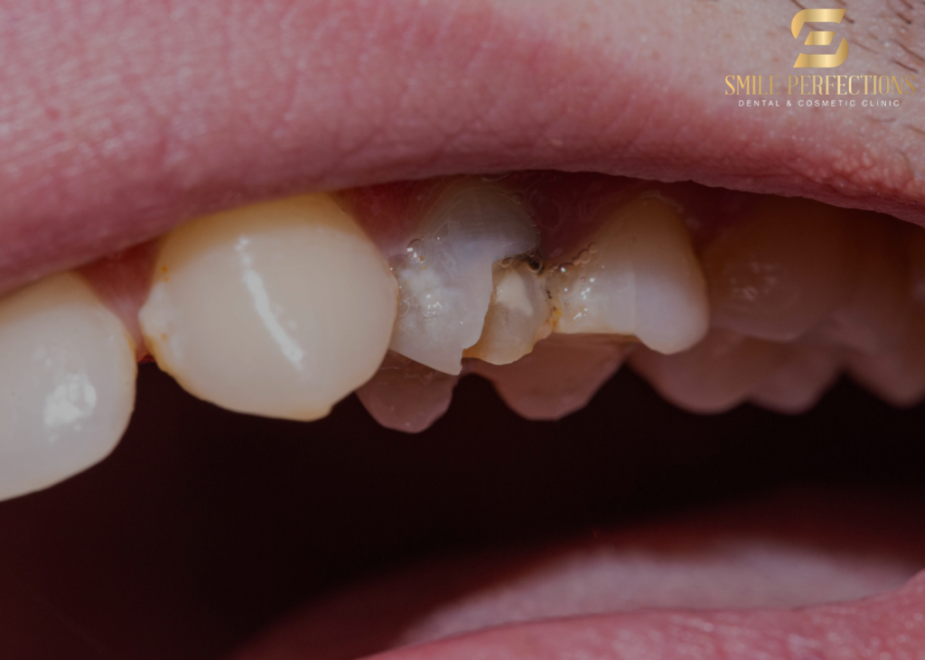
The fundamental distinction: intrinsic versus extrinsic discolouration
Every cause of a black tooth falls into one of two categories, and this distinction is the starting point for everything else.
- Extrinsic discolouration is on or within the outer surface of the tooth. The staining has originated from something outside the tooth, whether from food, drink, tobacco, bacteria or mineral deposits, and has adhered to or penetrated the enamel surface from the outside. Extrinsic staining ranges from light yellow-brown surface deposits to very dark, almost black staining in heavy tobacco or coffee users.
- Intrinsic discolouration originates from within the tooth structure itself. The colour change is in the dentine or pulp, not on the enamel surface. Intrinsic discolouration cannot be removed by cleaning or polishing, because it is not on the surface: it is in the body of the tooth. It typically requires either cosmetic coverage (composite bonding, veneers, crowns) or, in some cases, treatment of the underlying cause.
A tooth that looks black may have extrinsic staining, intrinsic discolouration, or both. The clinical approach to identifying which is straightforward: professional cleaning that removes surface deposits and then reassesses what remains. What is gone after cleaning was extrinsic. What persists is intrinsic.
Cause 1: dental decay — the most common cause of intrinsic blackening
Tooth decay is the most common reason for a tooth to turn black, and it is also one of the most clinically significant. Decay that has progressed through the enamel into the dentine changes the colour of the tooth structure it destroys: the bacterial acids demineralise the dentine, and the pigments produced by cariogenic bacteria turn the decayed tissue yellow, then brown, then black as the process advances.
Black decay in a tooth does not look the same as surface staining. It tends to be:
- Located in a specific site: the fissures on the biting surface, the contact between adjacent teeth, or around an existing filling
- Visible as a hole or cavity rather than a surface deposit
- Associated with sensitivity or, in more advanced cases, pain
- Sometimes accompanied by structural breakdown of the tooth (crumbling or fracture)
The blackness of advanced decay indicates significant bacterial metabolic activity within the tooth structure. This is not cosmetic: it requires removal of the decayed tissue and restoration of the cavity. Depending on the depth of the decay, this may involve a simple filling, a crown, or root canal treatment if the pulp is involved.
A dental check-up with X-rays is essential for establishing the extent of decay that has produced a black tooth: what is visible on the surface often represents only a fraction of the decay that has progressed laterally and internally beneath it.
Cause 2: pulp necrosis (a dead tooth)
A tooth that has turned dark grey, brown or black without any visible surface decay or staining has very likely undergone pulp necrosis: the internal pulp (nerve) tissue has died.
When the pulp dies, the blood vessels within it break down, and haemoglobin from red blood cells is released into the pulp chamber and then diffuses into the surrounding dentinal tubules. Haemoglobin breaks down into iron sulphide compounds that produce the characteristic dark grey to black discolouration of necrotic tooth tissue. The pigment permeates the dentine, producing discolouration that is visible through the enamel from outside.
What causes the pulp to die?
- Untreated decay that progressed to the pulp: the pulp becomes inflamed (pulpitis), then infected, and eventually necrotic
- Trauma: A blow to the tooth, whether from a fall, sport, or accident, can sever or damage the blood vessels entering through the root apex, cutting off the blood supply. The pulp dies without infection in these cases. The tooth may look completely intact externally while turning progressively darker over weeks to months after the injury
- Previous dental treatment: A very deep filling or repeated dental procedures on the same tooth can cause progressive pulp stress that eventually leads to necrosis
How a dead tooth presents: Grey-brown or black discolouration, often of a single tooth, sometimes preceded by pain and then followed by spontaneous resolution of pain (which is incorrectly interpreted as improvement but actually indicates the nerve has died). The tooth may be sensitive to percussion, and there may be a periapical abscess at the root tip producing a sinus tract (gum boil) on the adjacent gum.
Treatment: Root canal treatment removes the necrotic pulp, cleans and seals the canal system, and the tooth is then restored. The discolouration that remains after root canal treatment may be addressed with internal bleaching (whitening agent placed inside the pulp chamber), or with a crown or composite bonding to mask the remaining discolouration from outside.
Cause 3: old amalgam restorations casting a shadow
Amalgam, the silver-coloured metal filling material, has a distinctive effect on tooth appearance over time. As an amalgam filling ages, its metallic components — particularly the silver sulphide compounds that form as the material oxidises — diffuse into the surrounding dentinal tubules and cast a dark shadow through the overlying tooth structure.
This amalgam shadow appears as a dark grey or black discolouration visible through the tooth enamel, typically concentrated around an existing silver filling. The tooth itself is not decayed or damaged: the discolouration is caused by the metallic pigment that has migrated into the surrounding dentine.
This is both an aesthetic and a clinical concern. Aesthetically, the shadow makes the tooth look darker and unhealthy. Clinically, the appearance can sometimes obscure whether decay is also present, which is one reason X-rays are important at dental check-up appointments when large old amalgam fillings are present.
Treatment: Replacing the amalgam with a tooth-coloured composite or ceramic restoration removes the source of the metallic shadow. In some cases, composite bonding to the affected surfaces can visually address the remaining discolouration in the surrounding tooth structure. For more significant cases, a crown may be the most appropriate restoration.
Cause 4: extrinsic staining from tobacco, food and drink
The most visible extrinsic cause of a dark or black tooth is heavy tobacco use, particularly pipe smoking or chewing tobacco. Tobacco tar and nicotine compounds produce deep brown to black staining that adheres to the enamel surface and, over time, penetrates into the surface layers. This staining is difficult to remove with brushing alone and typically requires professional polishing and scaling to address.
Coffee, tea and red wine all contain chromogen compounds (colour-bearing molecules) and tannins that increase the permeability of the enamel surface to those chromogens. Heavy, frequent consumption over years produces brown to black staining that starts at the gum margins and in the fissures of the back teeth, where plaque provides a surface for the pigments to adhere to.
How to distinguish extrinsic from intrinsic staining: Extrinsic staining tends to be concentrated at the gum margin, in the fissures of the biting surface, and often distributes across multiple teeth rather than just one. It has a surface-like quality and can often be partially removed by professional cleaning.
Treatment: Professional cleaning and polishing at a dental hygienist appointment removes superficial and semi-superficial extrinsic staining. Air polishing in particular is highly effective for heavy tobacco and dietary staining. For staining that has penetrated the enamel surface and does not respond fully to polishing, teeth whitening or composite bonding on the affected surfaces can address what remains.
Cause 5: iron supplements
Liquid iron supplements, most commonly prescribed for iron deficiency anaemia, cause a characteristic black staining of the teeth that is superficial but very resistant to normal brushing. The iron reacts with sulphur compounds in saliva to produce iron sulphide, which adsorbs strongly to the tooth surface, producing dense black deposits, often concentrated at the gum line.
This staining is not evidence of decay or dental disease: it is a chemical reaction on the tooth surface. It is, however, persistent and very difficult to remove at home. Professional cleaning at a dental hygienist appointment can remove iron staining effectively in most cases.
Patients taking liquid iron can reduce the staining by diluting the supplement, drinking it through a straw to minimise contact with teeth, and rinsing thoroughly with water immediately after taking it.
Cause 6: black tartar (subgingival calculus)
While most tartar (calculus) is cream or yellow-white in colour, the tartar that forms below the gum line in the periodontal pockets of patients with gum disease takes on a characteristically dark brown to black appearance. This black or very dark tartar is called subgingival calculus.
Its dark colour comes from blood products in the gingival crevicular fluid and from the iron and other mineral compounds that incorporate into the calculus as it mineralises in the subgingival environment. When this calculus is close to the gum margin and becomes exposed through gum recession, it produces the appearance of a black band around the base of the tooth.
This is a clinical indicator of gum disease and bone loss, not just cosmetic staining. The appropriate response is periodontal assessment at a dental check-up and professional removal of the subgingival calculus at a dental hygienist appointment.
Cause 7: trauma to a developing tooth
In children, a significant blow to a primary (baby) tooth can cause haemorrhage within the pulp of the baby tooth, producing exactly the same grey-black discolouration mechanism as pulp necrosis in adults: blood breakdown products seep into the dentinal tubules and produce the characteristic discolouration.
A darkened baby tooth after a fall or blow does not always require extraction. If the tooth is not infected, not causing pain, and is not compromising the development of the permanent tooth beneath it, monitoring at regular dental check-up appointments may be the appropriate management. If infection develops, extraction becomes necessary.
The permanent tooth developing beneath the traumatised baby tooth can sometimes be affected by the injury, producing a discolouration, malformation or dilaceration (bending) of the developing permanent tooth. This is one of the reasons any significant dental trauma in childhood warrants professional assessment.
What to do if you notice a black tooth
The appropriate response depends on whether the blackening is extrinsic (on the surface) or intrinsic (within the tooth), and whether there are any associated symptoms.
- If there is pain alongside the black tooth: A black tooth with significant pain may indicate either deep decay reaching the pulp or an infected, necrotic tooth. This needs clinical assessment promptly, not observation at home.
- If the tooth is painless but progressively darkening: A single tooth becoming progressively darker without pain may have an ischaemic or post-traumatic pulp necrosis. It still needs assessment because the internal changes are progressing and an abscess can develop without pain if the nerve has died.
- If multiple teeth have surface dark deposits: This is most likely extrinsic staining from dietary, tobacco or tartar accumulation. A dental hygienist appointment addresses this directly. A dental check-up confirms whether any underlying decay is present.
- If a black tooth is treated but discolouration persists: Where the underlying cause has been managed but residual discolouration remains in the tooth structure, composite bonding can address the appearance. Composite applied to the labial (front) surface of a discoloured tooth, carefully matched to the surrounding teeth, masks the underlying discolouration and produces a natural result without requiring any irreversible tooth preparation in most cases.
The bottom line
A black tooth is not a single condition: it is a range of presentations from entirely superficial staining addressable with professional cleaning, to intrinsic discolouration from pulp necrosis requiring root canal treatment and restoration, to deep decay requiring immediate intervention.
What every black tooth has in common is that it needs clinical assessment at a dental check-up to establish the cause accurately before any treatment is planned. The treatment that addresses surface staining does nothing for a necrotic tooth. The treatment that addresses a necrotic tooth is not what is needed for an amalgam shadow.
At Smile Perfections in Oadby, Leicester, Dr Juttes Pallipatt GDC No. 104499 and Dr Pratima Pallipatt GDC No. 101258 assess discoloured teeth systematically, establish the specific cause, and recommend the most conservative and effective treatment path for each individual presentation.
Patients frequently ask
In most cases, yes, though the extent of treatment depends on the cause. A tooth darkened by pulp necrosis can be saved with root canal treatment to remove the necrotic tissue, followed by restoration to address both structure and appearance. A tooth darkened by deep decay can often be saved if enough sound structure remains above the gum line. Only where decay has destroyed the tooth below the gum line, where a vertical root fracture has occurred, or where bone loss is too severe to support treatment, is extraction the appropriate recommendation. A dental check-up with X-rays establishes exactly what is present.
Not always. A single black or darkened tooth is often caused by pulp necrosis following trauma rather than decay, particularly if there is no history of dental problems with that tooth. Black staining across multiple teeth is more likely extrinsic (from tobacco, coffee or iron supplements) than decay in most patients. Dark deposits visible at the gum line often indicate subgingival tartar rather than tooth decay itself. A thorough clinical assessment identifies which cause applies and prevents unnecessary treatment assumptions.
Yes, composite bonding is an effective option for addressing the appearance of a discoloured tooth once any underlying clinical cause has been treated. Composite resin applied to the front surface of a darkened tooth can mask the discolouration with a colour-matched, natural-looking layer. It requires no irreversible preparation of the tooth in most cases, is completed in a single appointment, and is reversible. For teeth with significant intrinsic discolouration from pulp necrosis or amalgam shadow, it can produce a transformation that significantly improves the smile without the need for more invasive restorations.
A tooth that darkens progressively after a blow, sometimes weeks or months after the original injury, has most likely undergone traumatic pulp necrosis. The blow damaged or severed the blood vessels entering through the root apex, cutting off the blood supply. As the pulp tissue broke down, haemoglobin from the blood was released and its breakdown products (iron sulphide compounds) diffused into the dentinal tubules, producing the characteristic grey-black discolouration. The tooth should be assessed at a dental check-up to confirm whether the pulp is non-vital and whether a periapical abscess has developed at the root tip.
For extrinsic black staining, a dental hygienist appointment involves professional scaling to remove any tartar (calculus) associated with the staining, followed by air polishing using a sodium bicarbonate powder system that removes surface staining very effectively. For tobacco or dietary staining that has penetrated slightly into the enamel surface, the hygienist may also use prophy paste polishing to restore the surface. If staining persists after professional cleaning, it is intrinsic rather than extrinsic, and a different approach is needed, which is discussed at the check-up stage.
Medical and dental information disclaimer
The information in this article is intended for general educational guidance only and does not constitute personalised dental advice. For concerns about a dark or discoloured tooth, please book an appointment with a qualified dental professional for a proper clinical assessment.
Smile Perfections is a private dental practice in Oadby, Leicester, led by Dr Juttes Pallipatt GDC No. 104499 and Dr Pratima Pallipatt GDC No. 101258. We offer dental check-ups, composite bonding, dental hygienist appointments, Invisalign, porcelain veneers, teeth whitening, dental crowns and smile makeovers.